Traumatic Optic Neuropathy

Traumatic Optic Neuropathy

Our vision plays a vital role in how we perceive and navigate the world. Following a significant head or facial injury, the delicate structures responsible for sight can be damaged, leading to serious visual impairment. Traumatic Optic Neuropathy is a condition caused by direct or indirect trauma to the optic nerve, often resulting in sudden and significant vision loss. This article explores its causes, symptoms, diagnostic approach, and the essential role of neuro-rehabilitation in supporting recovery and long-term visual function.

Traumatic Optic Neuropathy is a serious optic nerve injury caused by head or facial trauma that can lead to sudden vision loss. This guide explains its causes, symptoms, diagnosis, and how specialised neuro-rehabilitation helps patients adapt, recover function, and regain independence.

Understanding Traumatic Optic Neuropathy (TON)

To understand TON, one must first appreciate the intricate connection between the eye and the brain. This connection is not merely a simple wire but a complex, high-speed data cable that is vulnerable to injury.

The Critical Role of the Optic Nerve

The optic nerve is an incredible bundle of more than a million individual nerve fibers. Its sole purpose is to act as a communication superhighway, transmitting visual information captured by the retina at the back of the eye directly to the brain's visual cortex. The brain then interprets these signals, allowing us to perceive shapes, colours, and movement. Without a functioning optic nerve, sight is impossible, even with a perfectly healthy eye.

How Trauma Causes Optic Nerve Injury 

When the head or face sustains a powerful impact, the force can wreak havoc on the optic nerve. This can happen in several ways. Blunt or penetrating force may directly compress the nerve within its narrow bony channel (the optic canal), transect (cut) it, or disrupt the delicate blood vessels that supply it with oxygen and nutrients. When this supply is cut off, the nerve cells begin to malfunction and can eventually die, leading to irreversible vision loss.

Types of Traumatic Optic Neuropathy

Traumatic Optic Neuropathy is classified based on how the optic nerve is injured, which helps guide treatment decisions and prognosis.

Direct Traumatic Optic Neuropathy

This occurs when the optic nerve is physically damaged at the site of injury.

  • Caused by penetrating trauma or bone fragments from facial fractures
  • Often associated with visible orbital injury
  • Usually results in sudden and severe vision loss

Indirect Traumatic Optic Neuropathy

This is the more common form and occurs without direct injury to the eye socket.

  • Caused by the transmitted force from head trauma
  • The optic nerve is injured within the narrow optic canal
  • Damage occurs due to nerve stretching, swelling, or reduced blood flow

Causes of Traumatic Optic Neuropathy

Traumatic Optic Neuropathy occurs when a significant injury affects the optic nerve, usually following head or facial trauma. Common causes include:

  • High-impact head injuries: Road traffic accidents, major falls, or physical assaults that transmit force to the skull
  • Facial or orbital fractures: Broken bones near the eye socket or optic canal that compress or damage the optic nerve
  • Penetrating eye injuries: Trauma from sharp objects such as glass shards, metal fragments, or tools
  • Severe blunt force trauma: Sudden impact to the forehead or upper face causing shockwaves to travel to the optic nerve
  • Swelling or bleeding near the optic canal: Inflammation or hematoma that compromises blood supply to the nerve

Common Symptoms and Visual Field Defects

Traumatic Optic Neuropathy often presents with sudden and noticeable changes in vision shortly after head or facial trauma. These symptoms require immediate medical evaluation.

  • Sudden and severe vision loss: A rapid drop in vision in the affected eye, ranging from mild blurring to complete loss of light perception, often occurring immediately after the injury.
  • Abnormal pupil response (RAPD): When light is shone into the injured eye, the pupil reacts weakly.or not at all compared to the healthy eye, indicating impaired optic nerve function.
  • Visual field defects: Vision loss may affect specific areas rather than the entire visual field. Common patterns include central vision loss, making reading and face recognition difficult, or partial loss of peripheral vision.
  • Impaired colour vision: Colours may appear faded or washed out, with particular difficulty distinguishing shades of red, often one of the earliest signs of optic nerve damage.

Key Risk Factors for Developing TON

While any significant head trauma can potentially cause TON, certain scenarios and individuals are at a higher risk.

  • High-Impact Head or Facial Injuries: Individuals involved in motor vehicle accidents, significant falls from a height, or violent assaults are at the highest risk due to the immense forces exerted on the skull and orbital structures.
  • Participation in Contact Sports: Athletes in sports like boxing, mixed martial arts, and football are susceptible due to the frequency of direct blows to the head and face.
  • Pre-existing Cranial or Orbital Abnormalities: Some people are born with anatomical variations, such as an unusually narrow optic canal. In these individuals, even less severe trauma can cause significant nerve compression and damage.

The Diagnostic Process for Traumatic Neuropathy

Diagnosing Traumatic Optic Neuropathy (TON) requires prompt and detailed evaluation to assess optic nerve damage and rule out associated head or orbital injuries.

  • Comprehensive eye examination: An initial assessment measures visual acuity, pupil reactions, and colour vision to evaluate how severely the optic nerve has been affected.
  • Traumatic Optic Neuropathy fundus examination: Using an ophthalmoscope, the specialist examines the optic disc at the back of the eye. While it may appear normal early on, progressive pallor or atrophy over weeks confirms ongoing optic nerve damage.
  • CT and MRI scans: Imaging plays a critical role in diagnosis. CT scans help detect orbital or skull fractures, while MRI scans identify soft tissue injury, swelling, or blood clots that may be compressing the optic nerve and contributing to vision loss.

Differentiating TON from Other Post-Traumatic Neuropathies

Traumatic injuries can affect multiple nerves in the body. Understanding how Traumatic Optic Neuropathy (TON) differs from other post-traumatic nerve conditions is essential for accurate diagnosis and appropriate treatment.

Post-Traumatic Neuropathy: A Broad Term

Post-traumatic neuropathy refers to nerve damage that occurs anywhere in the body following physical injury. It can involve sensory, motor, or autonomic nerves. Traumatic Optic Neuropathy is a specific subtype, limited exclusively to the optic nerve and primarily affecting vision.

TON vs Post-Traumatic Trigeminal Neuropathy

Traumatic Optic Neuropathy should not be confused with post-traumatic trigeminal neuropathy. The trigeminal nerve controls facial sensation and jaw movement. Damage to this nerve typically causes:

  • Facial numbness or tingling
  • Sharp or burning facial pain
  • Difficulty chewing or jaw weakness

In contrast, TON affects visual pathways, leading to partial or complete vision loss without facial sensory symptoms.

A Key Clinical Difference: Pain

Pain helps differentiate these conditions. Although the injury causing TON may be painful, vision loss itself is usually painless. On the other hand, traumatic trigeminal neuropathy is often marked by persistent, severe neuropathic facial pain, making pain location and quality an important diagnostic clue.

Modern Treatment and Management Strategies

Managing Traumatic Optic Neuropathy focuses on minimising further optic nerve damage, reducing inflammation, and preserving any remaining vision. Treatment decisions are time-sensitive and depend on the severity of injury, imaging findings, and overall clinical presentation. Common management approaches include:

  • Medical Management (High-Dose Corticosteroids): Often administered intravenously soon after the injury, high-dose steroids aim to reduce inflammation and swelling around the optic nerve. Their effectiveness remains a topic of debate and is decided on a case-by-case basis.
  • Surgical Decompression: If imaging shows that the optic nerve is being compressed by bone fragments, a foreign body, or a hematoma, an emergency surgical procedure may be performed to decompress the optic canal and relieve the pressure.
  • Neuroprotective Agents and Future Therapies: Researchers are actively investigating new drugs and therapies that could protect nerve cells from dying after an injury and potentially promote regeneration, offering hope for future treatments.

Role of Neuro-Rehabilitation in TON Management

After the acute medical phase, neuro-rehabilitation plays a vital role in helping individuals adapt to vision changes, regain functional independence, and improve overall quality of life. Key components include:

  • Vision rehabilitation therapies: Focus on maximising remaining vision through techniques such as eccentric viewing and visual scanning, enabling safer navigation and improved daily functioning despite visual field loss.
  • Balance and coordination training: Addresses impaired depth perception and spatial awareness caused by vision loss, reducing fall risk and helping the body adapt by strengthening reliance on proprioceptive and vestibular inputs.
  • Adaptive and daily living skills: Occupational therapy supports practical independence by teaching modified techniques for everyday activities, recommending assistive tools, and helping patients adapt their home and work environments safely.

How to Care for a Loved One with Traumatic Optic Neuropathy

Supporting a loved one through the aftermath of TON requires patience, empathy, and practical adjustments.

  • Creating a Safe Home Environment: Simple changes can make a huge difference. Ensure rooms are well-lit, remove trip hazards like loose rugs, clear pathways of clutter, and use high-contrast tape to mark the edges of stairs.
  • Providing Emotional and Psychological Support: The sudden loss of vision can be emotionally devastating, leading to anxiety, depression, and a sense of isolation. Be a patient listener, offer encouragement, and help connect them with professional mental health support or peer groups.
  • Assisting with Appointments and Therapy: Navigating the healthcare system can be overwhelming. Offer to drive them to appointments, take notes during consultations, and help them practise their prescribed therapy exercises at home.

Factors to Consider When Choosing a Neuro Rehab Centre for TON

Selecting the right rehabilitation partner is a critical decision for patients and their families.

  • Multidisciplinary Team Approach: Look for a centre like Physiotattva that provides a cohesive team of physiotherapists, occupational therapists, and vision specialists who collaborate to create a holistic and comprehensive care plan.
  • Experience with Neurological and Vision Conditions: Ensure the facility has a proven track record of successfully treating patients with complex neurological injuries, especially those involving the visual system.
  • Personalized Treatment Plans: Recovery from TON is not a one-size-fits-all process. The best centres conduct a thorough initial assessment to create a customised rehabilitation program tailored to your specific visual deficits, functional limitations, and personal goals.

Personalised Care for Traumatic Optic Neuropathy Recovery at Physiotattva

Living with Traumatic Optic Neuropathy can be life-altering, but with the right care and support, meaningful recovery and adaptation are possible. Early diagnosis, timely medical intervention, and structured neuro-rehabilitation play a vital role in protecting function and improving long-term outcomes. With expert guidance, individuals can relearn daily skills, restore confidence, and regain independence. Our multidisciplinary neuro-rehabilitation team is dedicated to delivering personalised care tailored to your specific visual and functional needs.

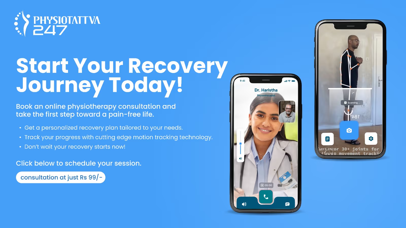
At Physiotattva physiotherapy clinics in Bangalore and Hyderabad, you receive personalised care tailored to your specific needs, ensuring effective results and comfort throughout your journey to recovery. 

Don’t wait to start your recovery! Get in touch with Physiotattva for more details! Contact us at +91 89510 47001.

Frequently Asked Questions

Can vision be fully restored after Traumatic Optic Neuropathy?

FAQ Icon
The prognosis varies widely. While some individuals experience a degree of spontaneous recovery, complete restoration is rare. Neuro rehabilitation focuses on maximizing the remaining vision and adapting to the changes.

Is Traumatic Optic Neuropathy the same as a stroke in the eye?

FAQ Icon
No. While both cause vision loss, TON is caused by physical trauma. An "eye stroke" (retinal artery occlusion) is typically caused by a blockage in the blood vessels supplying the retina due to cardiovascular issues.

How long does rehabilitation take for TON?

FAQ Icon
Rehab duration is highly individual. It depends on the severity of vision loss, the patient's goals, and their progress. It is an ongoing process of learning new skills and strategies.

What is the difference between traumatic optic neuropathy and post traumatic trigeminal neuropathy?

FAQ Icon
Traumatic optic neuropathy affects the optic nerve, causing vision loss. Post traumatic trigeminal neuropathy affects the trigeminal nerve, causing facial pain, numbness, or tingling.

Related Conditions

Related Treatments

No items found.

Related Symptoms

No items found.

Related Therapies

No items found.

Our Professionals

No items found.
No items found.
No items found.

Related Blogs

No posts published yet.

Get In Touch

Valid number
Thank you! Your submission has been received!
Oops! Something went wrong while submitting the form.